一 : 叩诊锤论坛moyamoaya氏病
典型影像竞猜

水货 昵称:

等级: 叩诊锤主治医师 发贴: 273 贴 总积分: 75 分 第27期 评奖积分: 0 分 注册: 2008-03-16
#1


2008-7-7 21:56:00


典型影像竞猜

查看此帖需要积分0 分


SRB521 版主 昵称:
等级: 叩诊锤主治医师 发贴: 1225 贴 总积分: 223 分 第27期 评奖积分: 0 分 注册: 2007-08-07
#2
2008-7-7 22:28:00

Re:典型影像竞猜
DAVF?
和这个类似
本贴已被 作者 于 2008-7-7 22:31:47 编辑过
查看此帖需要积分0 分


hammer 昵称:
等级: 叩诊锤主治医师 发贴: 372 贴 总积分: 95 分 第27期 评奖积分: 0 分 注册: 2007-10-13
#3
2008-7-8 10:52:00

Re:典型影像竞猜
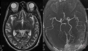
1.MoyaMoya:ivy征
http://www.61k.comforum/view.asp?id=3220
2.Sturge-Weber综合症
http://www.61k.comforum/view.asp?id=3901
3.DAVF
Quote: 以下是引用 yyzzhh 于 2008-6-4 20:17:39 的发言
引用其他朋友的一段话:
多发的点状血管流空影多见于以下几种情况:
A.以动脉为主的MOYAMOYA,以异常增生的小血管为主,但分布以双侧基底节为多。
B 动静脉均有的:
AVM——往往有明显的畸形血管团,其流空纵横交错,不呈散在点状分布。
DAVF——往往也流空纵横交错,部分只扫描到流空静脉,可呈散在点状分布,但以颅外供血为主往往有明显颅外血管流空,患者常有头痛头鸣史,头部有血管杂音,进一步行DSA检查明确。
C 以静脉为住:
静脉窦及皮层静脉血栓形成后相应引流区域皮层浅静脉迂曲扩张,在切面上呈散在点状分布流空影。该病人有发热史,可考虑上矢状窦血栓形成,但DSA肯定能明确。
另外还有脑静脉畸形:以扩张增多的髓静脉及粗大的引流静脉为主,在造影上呈典型水母头状,在MRI上亦有类似表现。
下面一例患者来源于其他论坛,最后的结论是硬脑膜动静脉漏。
本贴已被 作者 于 2008-7-8 10:56:25 编辑过
查看此帖需要积分0 分

神经新手 昵称:
等级: 叩诊锤住院总医师 发贴: 213 贴 总积分: 52 分 第27期 评奖积分: 0 分 注册: 2008-04-18
#4
2008-7-8 16:21:00
Re:典型影像竞猜
如果是硬脑膜动静脉漏(DAVF),如何解释患者的白质脑病?
查看此帖需要积分0 分

喋喋不休 昵称:
等级: 叩诊锤主治医师 发贴: 330 贴 总积分: 94 分 第27期 评奖积分: 0 分 注册: 2008-04-23
#5
2008-7-9 14:49:00

Re:典型影像竞猜
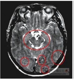
应该是典型的MoyaMoya病的ivy征(常青藤征)
看下面是以前收集的一个病例,几乎一样:
本贴已被 作者 于 2008-7-9 14:49:44 编辑过
查看此帖需要积分0 分

yyzzhh 管理员 昵称:忙!

等级: 叩诊锤主任医师 勋章:
发贴: 4720 贴 总积分: 701 分 第27期 评奖积分: 0 分 注册: 2007-03-30
#6
2008-7-10 21:27:00
Re:典型影像竞猜
白质脑病+T2相多发短T2异常信号(血管影,增粗的血管,静脉可能性大):硬脑膜动静脉瘘
下面是一个文献报道:
Dural arteriovenous fistula mimicking leukoencephalopathy.Neurology 2000;54:1123
A 49-year-old man had headache and fever for 1 month.Neurologic examination showed cognitive dysfunction: recent memory loss, dyscalculia, and disorientation. Muscle stretch reflexes were increased with Babinski signs. Sensory and cerebellar function were normal without meningeal signs. T2-weighted MRI revealed hyperintense signal areas in the internal capsules, globus pallidus, and subcortical white matter regions (A). Normal flow void appearance was not seen in the superior sagittal sinus (A, arrow).Diffusion-weighted imaging also disclosed diffuse hyperintensity in the subcortical white matter. In addition, T1-weighted MRI showed abnormal flow void sign in the cerebellum with gadolinium enhancement (B). Brain MR angiography suggested arteriovenous shunts. Arterial phase of the right external carotid arteriogram indicated dural arteriovenous fistula in the straight and transverse sinus (C). The venous phase of arteriogram demonstrated prominent venous collaterals and congestion in the great vein of Galen, inferior sagittal, and straight sinus (D). The internal carotid angiogram did not define the superior sagittal,transverse, and sigmoid sinus obviously. These images strongly support that perturbation of the venous outflow and sinus thrombosis could induce diffuse brain edema or infarction. Dural arteriovenous fistula occasionally causes a unique distribution of MRI lesions mimicking acute leukoencephalopathy.
图C提示:右侧颈外动脉直接和横窦和乙状窦相通
图D提示:直窦、下矢状窦和Galen静脉显影。
本贴已被 作者 于 2008-7-10 21:31:42 编辑过
查看此帖需要积分0 分
水货 昵称:
等级: 叩诊锤主治医师 发贴: 273 贴 总积分: 75 分 第27期 评奖积分: 0 分 注册: 2008-03-16
#7
2008-7-11 21:39:00
Re:典型影像竞猜
都是高手,的确就是硬脑膜动静脉瘘。
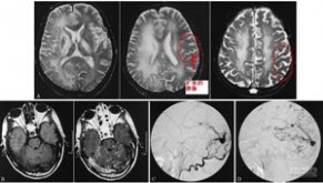
从MRI上可以发现很多血管的流空信号:
补充病史:A 67-year-old woman had in her medical history
an operation for an acoustic neurinoma on the right. She presented
with a rapid cognitive decline.The T2-weighted images
(first two rows) show a diffuse signal increase in the deep white
matter of both cerebral hemispheres.Abnormal flow voids are
visible on the cerebral surface and in the brain parenchyma,
especially in the temporal lobes and in the posterior fossa.The
T1-weighted images after contrast (third and fourth rows) show
extensive enhancement of cortical and parenchymal veins
公布DSA:
Angiography of the patient
presented in Fig. 101.2. Selective
injection of the external carotid artery
(first row, left) shows hypertrophy of
the superficial temporal artery and
middle meningeal artery. Immediate
enhancement of the superior sagittal
sinus is seen, suggesting the presence
of a DAVF. Selective internal carotid
artery injection (first row, right; middle
row, left) shows filling of the artery of
the falx cerebri through a (hypertrophied)
ophthalmic artery (first row,
right).There is early enhancement of
the superior sagittal sinus, consistent
with a DAVF.There is slow passage of
contrast through the brain parenchyma
because of venous congestion,
resulting in a late parenchymal and
venous phase.This is also illustrated
by the poor visibility of peripheral
arteries in the early phase (middle row,
left) and by the “pseudo-phlebitic”
aspect of the brain parenchyma in a
later phase of the angiogram (middle
row, right).After treatment by both an
endovascular approach (selective glue
injections in external carotid branches
feeding the arteriovenous fistula) and
by direct placement of coils in the
superior sagittal sinus (third row, left;
middle row, right), there is a marked
reduction in the size of external carotid
artery branches.The internal carotid
artery injection after treatment shows
abnormal drainage of the brain due to
occlusion of the superior sagittal sinus
(third row, right).The brain is no longer
using the superior sagittal sinus for its
drainage.However the transit time of
contrast is much shorter.The cognitive
symptoms of the patient have disappeared
almost completely
本贴已被 作者 于 2008-7-11 21:47:10 编辑过
查看此帖需要积分0 分

稀里糊涂 昵称:
等级: 叩诊锤住院总医师 发贴: 238 贴 总积分: 55 分 第27期 评奖积分: 0 分 注册: 2007-12-08
#8
2008-7-12 8:42:00
Re:典型影像竞猜
好病例,学习了。
查看此帖需要积分0 分


rose 超级版主 昵称:
等级: 叩诊锤主任医师 勋章:
发贴: 2865 贴 总积分: 672 分 第27期 评奖积分: 13 分 注册: 2008-08-11
#9
2009-6-1 22:11:00

Re:典型影像竞猜
ivy征一例:
360docimg_139_ 点击下载
本贴已被 作者 于 2009-6-1 22:12:43 编辑过
查看此帖需要积分0 分 360docimg_140_
二 : 叩诊锤论坛典型影像常春藤征(ivy sign)1
典型影像,大家看看

yyzzhh 管理员 昵称:忙!
等级: 叩诊锤主任医师 勋章:

发贴: 4720 贴 总积分: 701 分 第27期 评奖积分: 0 分 注册: 2007-03-30
#1


2008-1-25 14:49:00


典型影像,大家看看

A:平扫MRI T1
B:增强MRI T1
C:质子相
D:T2
查看此帖需要积分0 分

plateauhawk 昵称:

等级: 叩诊锤实习医师 发贴: 19 贴 总积分: 6 分 第27期 评奖积分: 0 分 注册: 2007-10-27
#2
2008-1-25 15:26:00
Re:典型影像,大家看看
什么东东?脑囊虫!!!!!!!
查看此帖需要积分0 分
yyzzhh 管理员 昵称:忙!
等级: 叩诊锤主任医师 勋章:
发贴: 4720 贴 总积分: 701 分 第27期 评奖积分: 0 分 注册: 2007-03-30
#3
2008-1-25 16:02:00
Re:典型影像,大家看看
上面的图片不清楚,发另一个病人的MRI增强片:

本贴已被 作者 于 2008-1-25 16:2:45 编辑过
查看此帖需要积分0 分

小不点120 昵称:
等级: 叩诊锤实习医师 发贴: 8 贴 总积分: 1 分 第27期 评奖积分: 0 分 注册: 2008-01-24
#4
2008-1-25 16:31:00
Re:典型影像,大家看看
脑沟加深,加增.脑白质见星粒改变.应该是"脑囊虫"吧
查看此帖需要积分0 分
xie780804 昵称:青囊行者
等级: 叩诊锤实习医师 发贴: 10 贴 总积分: 3 分 第27期 评奖积分: 0 分 注册: 2008-01-16
#5
2008-1-25 17:12:00
Re:典型影像,大家看看
强烈要求提供病史、查体资料!
查看此帖需要积分0 分

w88w 昵称:
等级: 叩诊锤副主任医师 发贴: 439 贴 总积分: 120 分 第27期 评奖积分: 0 分 注册: 2007-11-13
#6
2008-1-25 21:32:00
Re:典型影像,大家看看
增强扫描可以发现软脑膜增强,提示炎性疾病或者脑膜癌。这些不好解释患者脑室周围缺血改变。
脑膜炎?
血管炎?
脑膜癌?
查看此帖需要积分0 分
yyzzhh 管理员 昵称:忙!
等级: 叩诊锤主任医师 勋章:
发贴: 4720 贴 总积分: 701 分 第27期 评奖积分: 0 分 注册: 2007-03-30
#7
2008-1-26 13:03:00

Re:典型影像,大家看看
患者术中的图片:
本贴已被 作者 于 2008-1-26 13:4:4 编辑过
查看此帖需要积分0 分

chinastroke 名誉版主 昵称:I'm Wall.E
等级: 叩诊锤主治医师 发贴: 1961 贴 总积分: 232 分 第27期 评奖积分: 0 分 注册: 2007-06-30
#8
2008-1-26 13:08:00
Re:典型影像,大家看看
Quote: 以下是引用 yyzzhh 于 2008-1-26 13:03:24 的发言
患者术中的图片:
我明白了,软脑膜强化有一种原因为分布在软脑膜上的血管显著增多,这样造影剂在小血管中出现潴留,因此可看到软脑膜强化的情况。
脑膜癌的软脑膜强化和moyamoya的软脑膜强化机理是不是一样的呢?
这是不是也说明只要存在严重的软脑膜血管增生就会出现软脑膜强化呢?
Sorry!
永远支持论坛!
查看此帖需要积分0 分

zxp2005282 昵称:
等级: 叩诊锤住院医师 发贴: 94 贴 总积分: 30 分 第27期 评奖积分: 0 分 注册: 2007-06-29
#9
2008-1-26 16:15:00

Re:典型影像,大家看看
I got it
"Ivy Sign" in Moyamoya Disease.注:常春藤征(ivy sign)
look!
点击下载
and
点击下载
本贴已被 作者 于 2008-1-26 16:47:16 编辑过
查看此帖需要积分0 分
yyzzhh 管理员 昵称:忙!
等级: 叩诊锤主任医师 勋章:
发贴: 4720 贴 总积分: 701 分 第27期 评奖积分: 0 分 注册: 2007-03-30
#10
2008-1-26 20:02:00

Re:典型影像,大家看看
哈,的确是典型的ivy sign
公布答案:MoyaMoya病
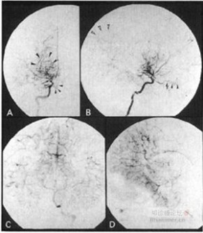
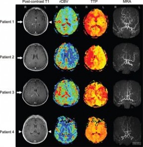
Figure 1. MR images of Patient 1 before the operation. A, precontrast T1-WI (TR, 400 ms; TE, 20 ms; 1 NEX). Several flow-void spots are seen corresponding to some cortical vessels (arrows). B, postcontrast T1-WI with Gd-DTPA (TR, 400 ms; TE, 20 ms; 1 NEX), showing diffuse leptomeningeal enhancement along cortical sulci, like ivy creeping between stones. The flow-voids seen in A remained almost the same (arrows). C, proton density image (TR, 2000 ms; TE, 30 ms; 1 NEX). Multiple flow-voids are seen in basal ganglia. D, T2-WI (TR, 2000 ms; TE, 90 ms; 1 NEX) shows subcortical highintensity lesions.

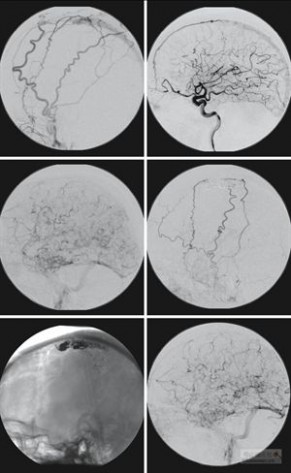
Figure 2. Carotid angiograms of Patient 1 before the operation. A and B, right carotid angiograms showing narrowing at the terminal portion of the internal carotid artery (arrow), basal moyamoya in evolution (large arrowheads), ethmoidal moyamoya in early stage (small arrowheads), parietal cortical arteries visualized via leptomeningeal anastomosis from posterior cerebral artery (open arrowheads). Cortical branches of the ACA and the MCA are poorly visualized. C and D, left vertebral angiograms showing marked retrograde filling into the territory of ACA and MCA via leptomeningeal anastomosis

Figure 3. Intraoperative view of Patient 1. Markedly profuse pial arterial network diffusely covers the cortex.
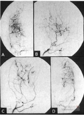
Figure 4. MR images of Patient 1 after the operation. A, precontrast T1-WI (TR, 400 ms; TE, 20 ms; 1 NEX) showing subtle increase in the cortical flow-void spots compared with Fig. 1A. B, postcontrast T1-WI with Gd-DTPA (TR, 400 ms; TE, 20 ms; 1 NEX) showing significant reduction in the leptomeningeal enhancement observed in Fig. 1B. C, proton density image (TR, 2000 ms; TE, 30 ms; 1 NEX). Multiple flow-voids in basal ganglia decrease compared with Fig. 1C. D, T2-WI (TR, 2000 ms; TE, 90 ms; 1 NEX) showing decrease in the subcortical high-intensity lesions.
360docimg_156_
Figure 5. Postoperative angiogram of Patient 1. A and B, right external carotid angiogram. Branches of MCA are opacified via surgical and leptomeningeal anastomosis. C and D, left external carotid angiogram. Similar findings as in A and B.
本贴已被 作者 于 2008-1-26 20:7:2 编辑过
本文标题:
叩诊锤论坛-叩诊锤论坛moyamoaya氏病 本文地址:
http://www.61k.com/1076982.html